
X-Ray Imaging
X-rays gave dental professionals a way to examine structures that could not be evaluated through visual examination alone.
The development of modern dentistry has been shaped by major advances in diagnostic imaging, restorative materials, laser technology and computer-aided dental systems. From the arrival of X-ray imaging to the development of CAD/CAM restorations, these innovations helped transform dental diagnosis, treatment planning and patient care.
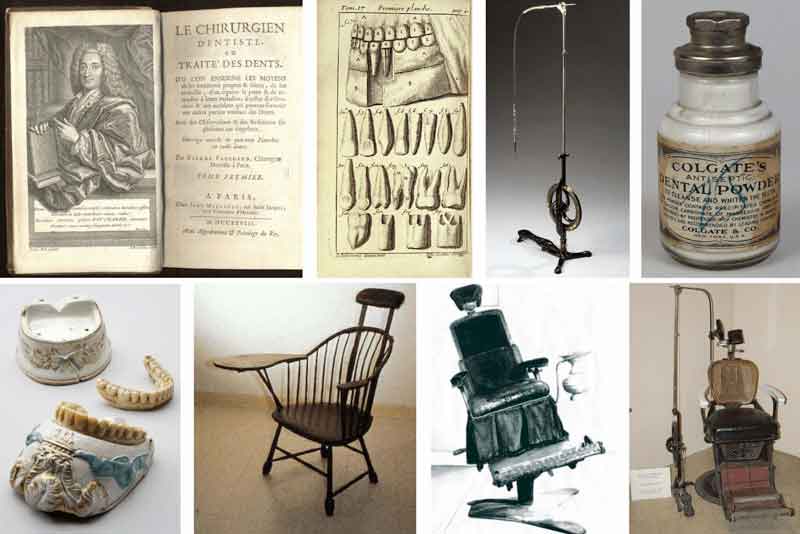
Dentistry changed considerably as new technologies expanded what dental professionals could see, diagnose, plan and restore.
Diagnostic imaging allowed structures beneath the visible surface to be evaluated. New restorative materials improved aesthetic possibilities, while laser systems introduced additional methods for selected dental procedures.
Computer-aided design and manufacturing later added a digital dimension to restorative dentistry by supporting the design and production of custom dental restorations.
The existing content highlights diagnostic imaging, restorative materials, laser dentistry and computer-aided systems as important developments in the evolution of dental care.
X-rays gave dental professionals a way to examine structures that could not be evaluated through visual examination alone.
Panoramic radiography expanded the ability to view broader dental and facial structures in a single image.
Tooth-colored restorative materials increased aesthetic options and introduced new bonding possibilities.
Dental lasers introduced additional precision-focused approaches for selected hard- and soft-tissue procedures.
Computer-aided systems enabled digital design and manufacturing of customised dental restorations.
The discovery of X-rays represented a major change in medical and dental diagnosis. Wilhelm Conrad Röntgen's work in 1895 introduced X-ray imaging, which was later incorporated into dentistry.
Dental X-rays allowed clinicians to evaluate teeth, bone and surrounding structures that were not directly visible during a routine examination.
This ability expanded diagnostic possibilities and became an important foundation for later developments in dental imaging technology.
Dental imaging continued to develop beyond conventional X-rays, introducing broader views and eventually three-dimensional information.
Radiographic imaging provided a way to evaluate dental and supporting structures that could not be seen through direct visual examination.
Panoramic imaging provides a wider view of the oral and maxillofacial region and can assist in assessing multiple dental and facial structures together.
Cone-beam computed tomography brought three-dimensional imaging into dental practice, supporting detailed assessment for applications such as implant planning and orthodontic evaluation.
Restorative dentistry also changed as dental materials developed. Traditional amalgam fillings were widely used but were visually noticeable because of their metallic appearance.
Tooth-colored composite materials introduced a more natural-looking restorative option and expanded the possibilities for adhesive dentistry.
The existing content highlights the development of tooth-colored composites as an important advancement because these materials improved aesthetic possibilities while also providing bonding capabilities that could support more conservative restorative approaches.
Laser technology became another important development in dentistry, introducing new tools for selected dental procedures.
The existing content identifies applications including cavity preparation, soft-tissue management and teeth whitening, reflecting the broader move toward more precise treatment techniques.
Computer-Aided Design and Computer-Aided Manufacturing introduced digital methods for designing and producing customised dental restorations.
CAD/CAM systems can be used in the production of restorations such as crowns, bridges and veneers, connecting digital design with restorative dentistry.
Imaging technologies made it possible to evaluate dental structures that could not be directly seen.
Panoramic and three-dimensional imaging expanded the information available for treatment assessment.
New dental materials increased aesthetic and adhesive possibilities in restorative dentistry.
CAD/CAM systems connected computer-based design with the manufacture of customised dental restorations.
Wilhelm Conrad Röntgen's discovery opened the way for radiographic imaging and its later use in dental diagnosis.
Dental radiography continued to develop and new imaging methods provided broader and more detailed views of dental structures.
Tooth-colored composite materials increased aesthetic restorative options and supported new adhesive techniques.
Digital imaging and computer-aided restorative systems expanded the role of technology in modern treatment planning and restorative dentistry.
The development of dental X-rays, panoramic and three-dimensional imaging, tooth-colored restorative materials, laser technology and CAD/CAM systems helped transform dentistry. Together, these advances expanded diagnostic capabilities, restorative options and digital treatment workflows and continue to influence the way modern dentistry is practised today.
Learn more about the dental technology used today or contact our team if you would like information about treatments and planning your dental care.